Onion Cell Diagram
Magnified microscope cell 40x microscopy micrographs walls Beautiful world: onion cells Onion cells
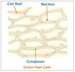
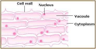
Onion Epidermal Cell Diagram
Cebola zwiebel microscope cipolla photomicrograph micrografia micrografo mikrograph resolution microscopio pino legno Onion epidermal cell diagram Onion peel cell diagram with label
Onion cell cells diagram structure 2010 biology microscopic occupies september uses introduction
Onion cells under microscopeOnion cells The science scoop: onion cell labOnion cells beautiful world.
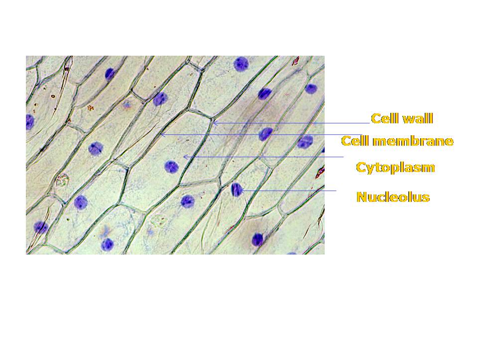
Onion peel cell diagram with labelOnion cell epidermal diagram labeled cells microscope under drawing skin epidermis lab bulb mag membrane observation vacuole nucleus leaves preparation Onion cells 2Onion epidermal drawings epidermis labeled biology chromosomes chromosome dna observation.

Onion mount temporary cells labelled draw cell prepare peel diagrams blissful earth objective observations record
Biology help online: september 2010Onion epidermal cell labeled diagram Onion cell epidermal peel sizeLab slides. cell types.
Onion microscope magnified 40x 100x microscopyOnion peel px Onion cells under microscopeLabel cells procedure.

Onion cell diagram drawing
Onion peel cell cells skin showing figure patternity savedOnion cell 400x lab microscope under labeled cells structure scoop science looked Biopedia: practicalsBlissful earth.
Epidermal epidermisOnion cell hi-res stock photography and images Figure of onion peel showing cellOnion cells cell lab types slides.
Onion cell micrograph microscope cells stock microscopic section root cepa allium scale alamy epidermis bulb tip organelles
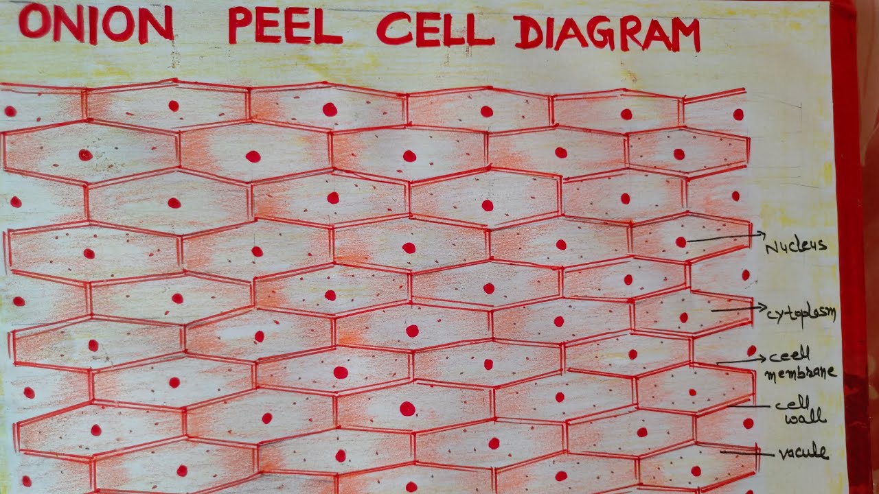
Cell peel ncertOnion epidermal cell diagram .
.
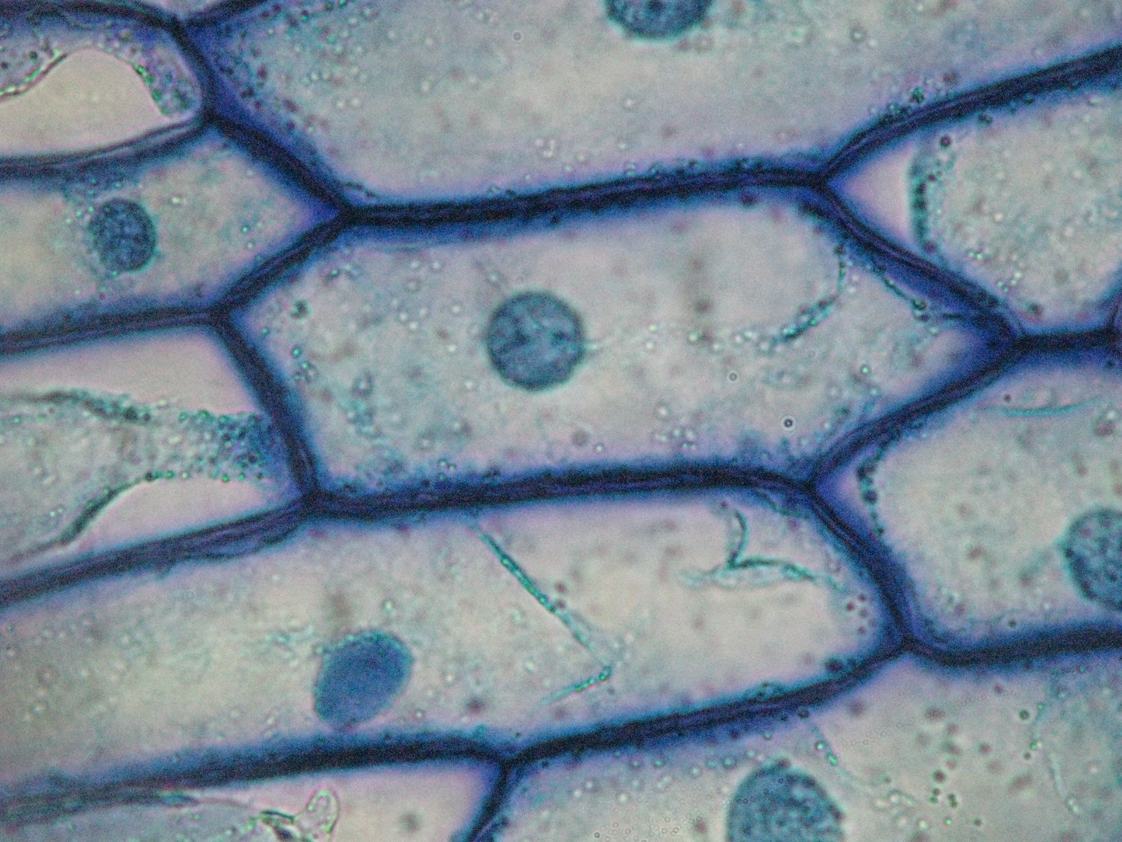
Beautiful World: Onion cells
Onion Cell Diagram Drawing - lana1970

Onion Cells under Microscope

Onion Peel Cell Diagram With Label - itsessiii
Blissful Earth

Figure of onion peel showing cell - Brainly.in

Onion Cells under Microscope

Onion cell hi-res stock photography and images - Alamy